Figure 1.
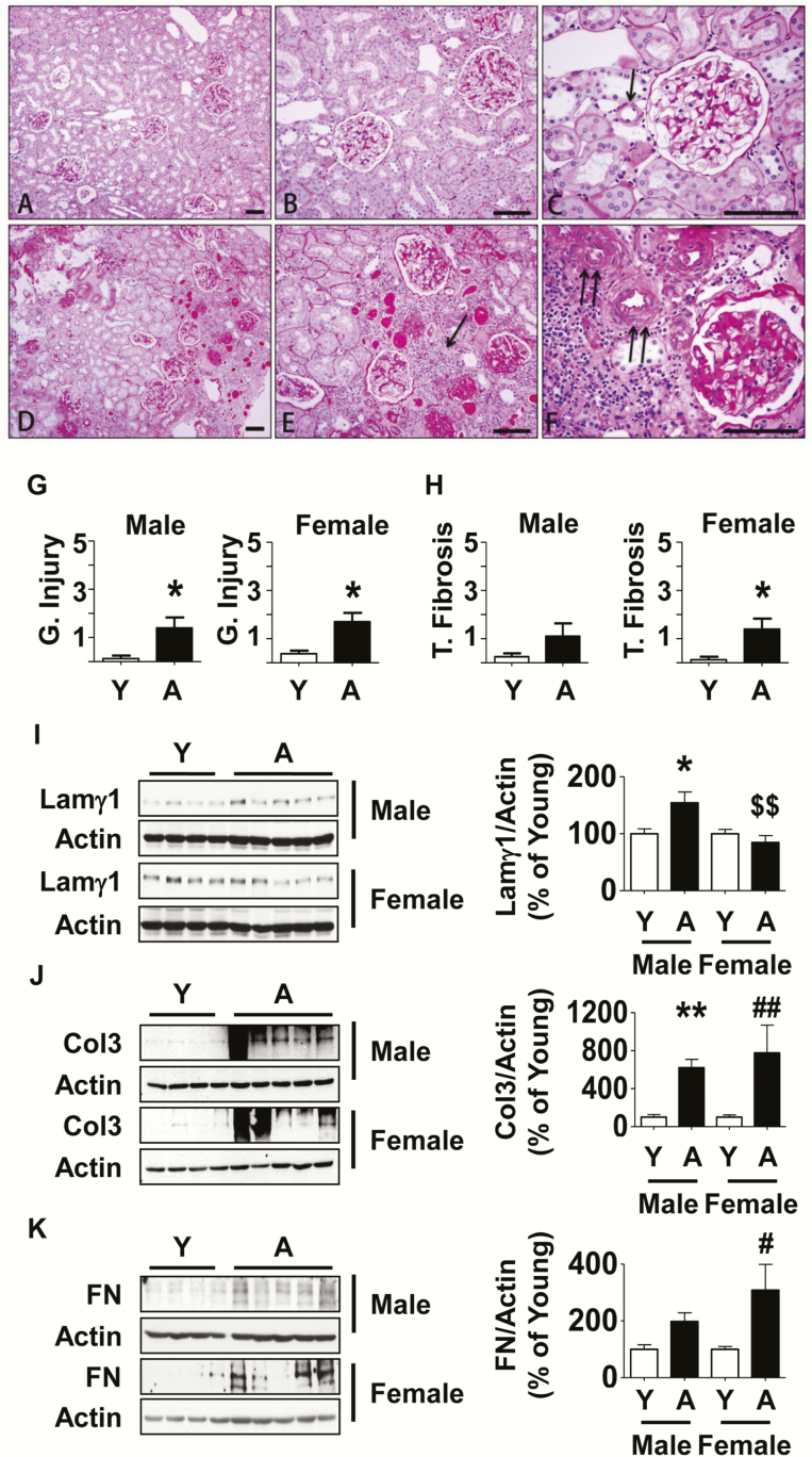
Kidney histology and extracellular matrix protein expression in aged marmosets. (A–C) By PAS stain, in young marmosets glomeruli displayed normal cellularity without segmental or global accentuation of mesangial matrix. Tubules were closely packed without atrophy. Blood vessels (arrow in C) were thin walled and lumens were patent. (D–F) In aged marmosets, glomeruli showed either segmental (part of an individual glomerulus) or total (whole of an individual glomerulus) sclerosis. Parietal glomerular basement membrane showed thickening and wrinkling. Tubules showed significant atrophy, loss, thickening of tubular basement membrane and many contained casts. There were rare areas of interstitial nephritis composed of mononuclear cells (arrow in E). Blood vessels showed narrowing of lumen and severe medial hypertrophy and sclerosis (double arrows in F). (G and H) Semi-quantitative scoring of indices of glomerular injury and tubular fibrosis are shown (Y = young, A = aged). Data are shown as mean ± SEM, n = 4 young male and n = 4 young female marmosets, n = 5 aged male and n = 5 aged female marmosets (*p < .05 by t-test). Renal cortical lysates were employed to perform immunoblotting using antibodies against (I) laminin γ1, (J) collagen III, and (K) fibronectin. Actin expression was used to assess loading (Y = young, A = aged). Data are shown as mean ± SEM, n = 4 young male and n = 4 young female marmosets, n = 5 aged male and n = 5 aged female marmosets (*p < .05, **p < .01 vs young male; #p < .05, ##p < .01 vs young female, $$p < .01 vs aged male by two-way ANOVA).

