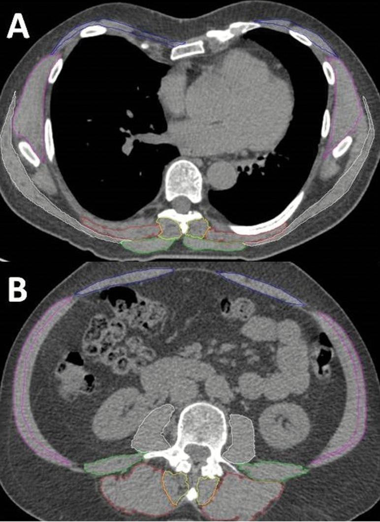
Figure 1.
Axial computed tomography images of the thoracic (A) and lumbar (B) muscles segmented using SpineAnalyzer. Thoracic muscles (A) measured at the midvertebral slice of T8 were the trapezius, erector spinae, and transversospinalis. Lumbar muscles (B) measured at the midvertebral slice of L3 were the erector spinae, transversospinalis, psoas major, quadratus lumborum internal, external obliques, and rectus abdominis.

