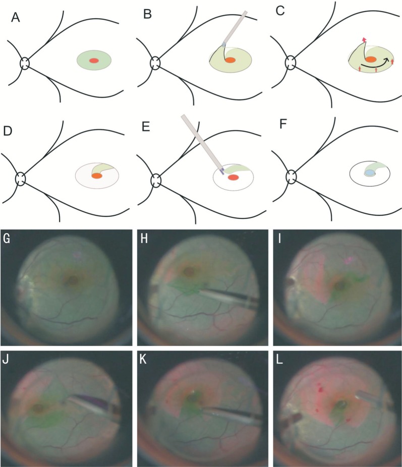
Figure 1. Diagrammatic representation of the tiled transplantation ILM pedicle flap technique and surgical video screenshot of one patient's left eye.
A, G: We stained the ILM with a 0.125% solution of ICG; B-D and H-K: The ILM was grasped and peeled off in a circumferential pattern for about 2-3 disk diameter around the MH using ILM forceps, leaving a pedunculated ILM flap; Peel the ILM in the direction of the red arrows; E, L: Then the ILM pedicle flap was tiled over the MH, and was carefully flattened to make sure it was properly positioned; F: Sometimes we placed a sodium chondroitin sulfate-sodium hyaluronate on the ILM flap to stabilize it.

