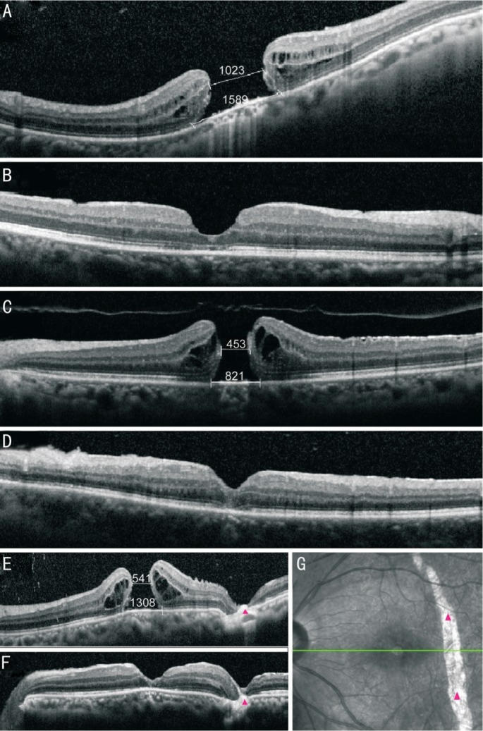
Figure 3. Spectral-domain OCT changes in three representative patients after PPV with the tiled transplantation ILM pedicle flap technique.

A, C, E: Preoperative OCT images; B, D, F: Seven days postoperative OCT images. Seven days postoperative, the MH had closed. The closure types of first patient was U-type and the other two patients were V-type. The third patient (E-G) is a 19-year-old female patient whose left eye was injured by a beer bottle. G: The fundus photography taken during OCT scanning. The trauma not only caused a large MH but also caused a choroidal rupture (position of the red arrow in the E-G). The MH healed successfully after the PPV with the tiled transplantation ILM pedicle flap technique. Vision increased from 20/500 to 20/40.
