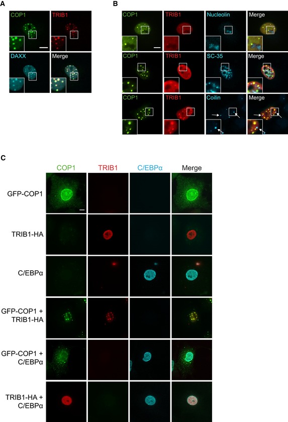
Figure EV2. COP1 and TRIB1 colocalize in punctate nuclear structures.

- Representative images of COS7 cells showing that COP1‐3xFLAG (green) and TRIB1‐HA (red) colocalize with GFP‐DAXX (cyan). Insets show enlarged images of nuclear puncta. Cells were stained with anti‐FLAG (Cell Signaling), anti‐HA (Santa Cruz), anti‐mouse Alexa Fluor 405, and anti‐rabbit Alexa Fluor 680. Scale bar, 10 μm.
- Representative images of COS7 cells showing that GFP‐COP1 (green) and TRIB1‐FLAG (red) do not colocalize with markers (cyan) for nucleoli (nucleolin), nuclear speckles (SC‐35), or Cajal bodies (coilin). Cells were stained with anti‐FLAG (Cell Signaling), anti‐mouse Alexa Fluor 405, anti‐rabbit Alexa Fluor 680, and either anti‐nucleolin, anti‐SC‐35, or anti‐coilin, as indicated. Scale bar, 10 μm.
- Representative images of COS7 cells expressing the indicated combinations of GFP‐COP1 (green), TRIB1‐HA (red), and C/EBPα (cyan). Images for cells coexpressing all three constructs are shown in Fig 1D. Cells were stained with anti‐HA (Santa Cruz), anti‐C/EBPα (Cell Signaling), anti‐mouse Alexa Fluor 405, and anti‐rabbit Alexa Fluor 680. Scale bar, 10 μm.
