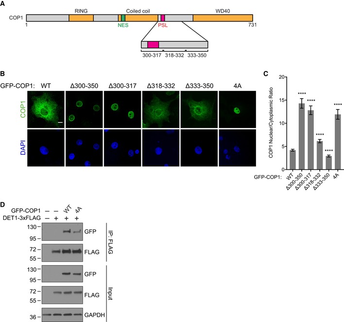
Figure EV5. Identification of an intramolecular WD40‐binding site that regulates the subcellular distribution of COP1.

- Schematic of COP1 illustrating locations of the coiled coil–WD40 domain linker deletions and PSL.
- Representative images of COS7 cells transfected with the indicated GFP‐COP1 constructs (green). Nuclei were counterstained with DAPI (blue). Scale bar, 10 μm.
- Quantification of the average ratio for nuclear/cytoplasmic fluorescence for each of the indicated GFP‐COP1 constructs. Mean values ± s.e.m. are shown for three independent experiments where 50 individual cells per experiment were analyzed. Significance relative to GFP‐COP1 WT was calculated using the Student's t‐test (****P < 0.00001).
- Coimmunoprecipitation of GFP‐COP1 with DET1‐3xFLAG in COS7 cells showing that the 4A mutation weakens the DET1/COP1 interaction.
