
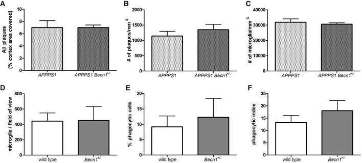
Figure EV2. Abeta pathology and phagocytic capacity are not affected by BECN1 reduction.
-
A–CStereological analysis of the cortex of 4‐month‐old APPPS1 and APPPS1‐Becn1 +/− mice was performed to assess the area covered by 4G8 (A), the number of core plaques stained with Congo red (B), and the number of Iba1+ microglia (C). Mean ± SEM, APPPS1 n = 4, APPPS1‐Becn1 +/− n = 2, two‐tailed t‐test ns.
-
D–FThe phagocytosis assay performed in acute brain slices of 4‐month‐old wild‐type and Becn1 +/− mice was assessed for the number of Iba1+ microglia (D), the percentage of phagocytic cells (E), and the phagocytic index (F). Mean ± SEM, wild‐type n = 5, Becn1 +/− n = 4, two‐tailed t‐test ns.