
Figure EV3. Decreased autophagy affect levels of activated CASP1, but not IL‐1beta or CASP1 precursors.
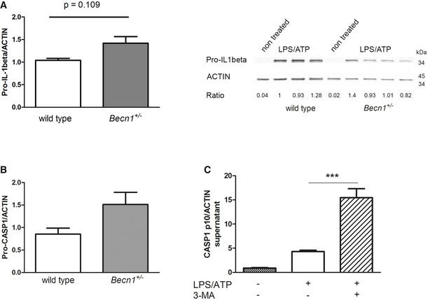
- Pro‐IL‐1beta in the cell lysates of LPS/ATP‐treated wild‐type and Becn1 +/− microglia was detected by Western blot. No significant differences can be detected between wild‐type and Becn1 +/− microglia; mean ± SEM, wild‐type LPS/ATP n = 11, Becn1 +/− LPS/ATP n = 26, two‐tailed t‐test ns.
- Pro‐CASP1 in LPS/ATP‐treated wild‐type and Becn1 +/− microglia was detected by Western blot in the cell lysates. No significant differences can be detected between wild‐type and Becn1 +/− microglia; mean ± SEM, wild‐type LPS/ATP n = 6, Becn1 +/− LPS/ATP n = 14; two‐tailed t‐test ns.
- Microglia were isolated from newborn wild‐type mouse pups and were either non treated or treated with a pro‐inflammatory stimulus (LPS followed by ATP) in the presence or absence of 3‐MA. The presence of cleaved CASP1 (p10) protein in the cell supernatant was determined by Western blot and normalized to ACTIN. Treatment with 3‐MA significantly enhanced the release of CASP1; mean ± SEM, n = 3, ANOVA with Tukey's post hoc test; ***P < 0.001.
Source data are available online for this figure.