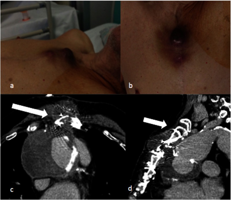
Fig. 1.
Picture of an 85-year-old man, presenting with a non-pulsatile swelling cutaneous lesion in the upper third of the sternal wound. The lesion was surrounded by a bruise area (a, b). CECT angiography scan revealed a large pseudoaneurysm measuring 70 mm in diameter arising from the distal suture line of the Dacron graft with a defect in the aortic wall of 5 mm and large parietal thrombus apposition (c, d). In addition, the sternum appeared with mild diastasis (arrow) and the pseudoaneurysm was abutting the sternum and extending through the bone to subcutaneous tissue. Anteriorly to the sternum was observed a fluid collection of 43 mm in connection with the pseudoaneurysm sac

