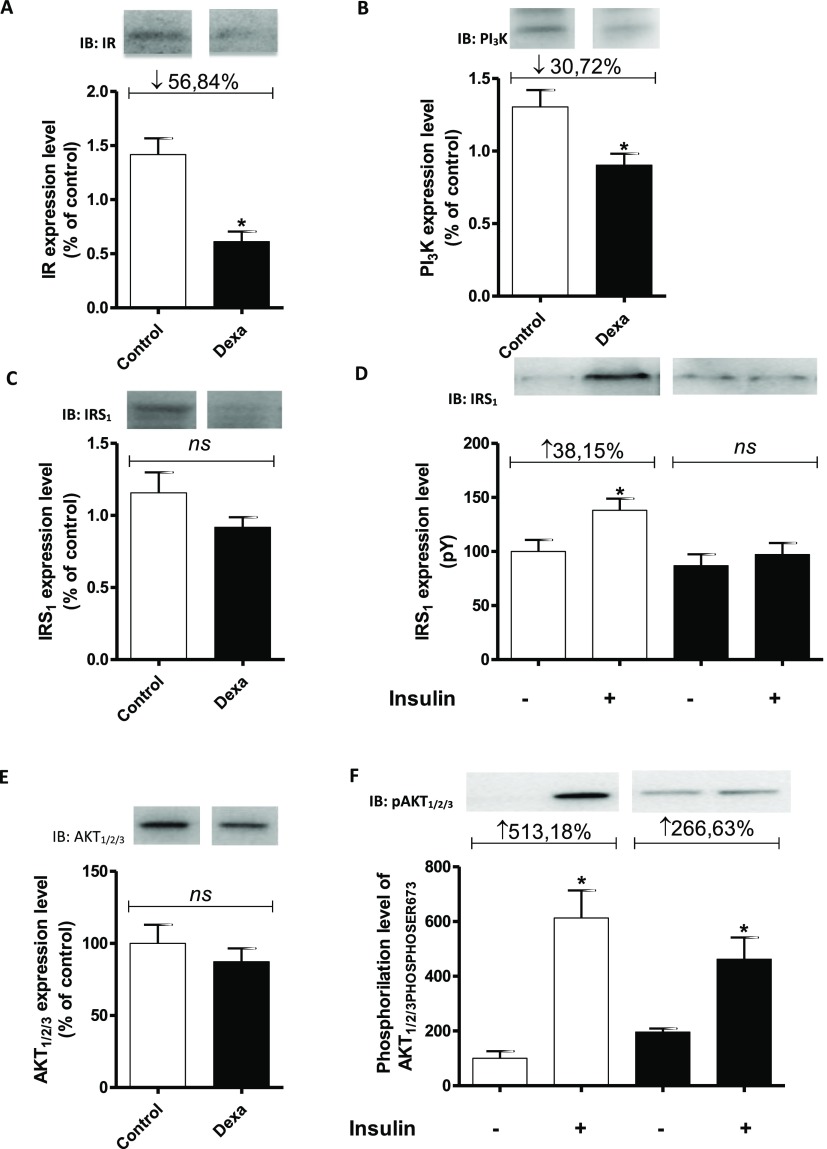
Fig. 2.
Samples containing 25–50 mg of solubilized proteins were subjected to SDS-PAGE and immunoblotting using specific antibodies. A blot representative of the experiments is shown. The status of phosphorylation and protein expression (percentage) involved in intracellular insulin signaling in the masseter muscle of rats in the CON (hollow bars; n = 6) and DEX groups (solid bars; n = 6) was determined by stoichiometry. Analysis of the degree of expression of IR (A), PI3K (B), and IRS1 (C) in the masseter muscle in the CON and DEX groups. Analysis of the degree of IRS1 phosphorylation/activity in the masseter muscle in the CON and DEX groups before [time zero (−)] and after the infusion of insulin into the portal vein (+) (D). Total amount of Akt protein in the masseter muscle in the CON and DEX groups (E). Analysis of the degree of phosphorylation/activity (phosphorylation at serine 473) of Akt in the masseter muscle in the CON and DEX groups before [time zero (−)] and after the infusion of insulin into the portal vein (+) (F). Student’s t-test was used in the intergroup analysis (*p < 0.05)

