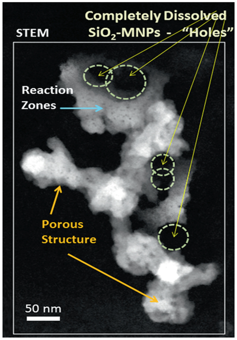
Fig. 4.5.
Dark field STEM imaging of lung section after repeated dose inhalation and 27 days post treatment. SiO2-MNPs show pores and significant in vivo processing. Almost all of the original spherical morphology has disappeared after 27 days post treatment exposure. SiO2-MNPs show dissolution patterns, void/pore formation (yellow arrows) and significant outward growth of reaction zones (secondary growth shown by blue arrow)

