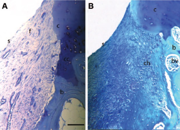
Figure 1.

Toluidine blue stained histology sections of the defect edge at (A) 1 week and (B) 4 weeks postinjury. In both A and B there is loss of proteoglycan at the cartilage edge and a loss of the normal architecture of the cartilage. Chondrocyte clusters are seen in B. In A, fibrous tissue fills the defect with a defined surface layer. In B the fibrous tissue filling the lesion is being replaced by rounded chondrocytes. c = cartilage edge; f = fibrous tissue in defect; b = bone; s = surface of tissue infill in defect; cc = calcified cartilage; ch = chondrocytes in defect; bv = blood vessel. Bar = 750 µm.
