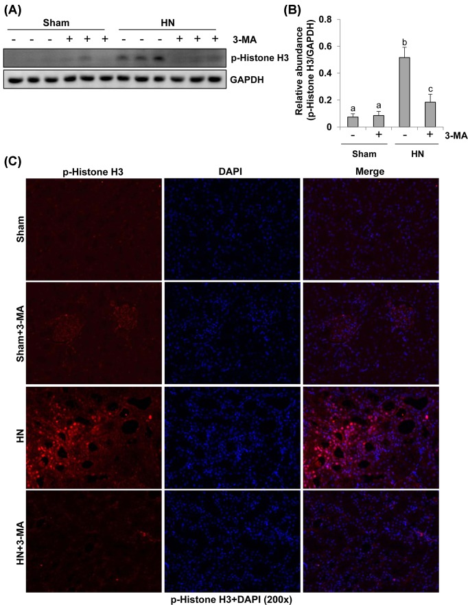
Figure 7. Administration of 3-MA reduces expression of p-histone H3 in hyperuricemic rats.
The prepared tissue lysates from sham or hyperuricemic kidneys of rats treated with/without 3-MA were subjected to immunoblot analysis with specific antibodies against p-histone H3 and GAPDH (A). The expression levels of p-histone H3 were quantitated by densitometry and normalized with GADPH (B). Photomicrographs (200×) illustrate p-histone H3 (Ser10) with immunofluorescent staining of the kidney tissues from sham or hyperuricemic kidneys of rats treated with/without 3-MA (C). Data are represented as the mean ± S.E.M. (n=6). Means with different superscript letters are significantly different from one another (P<0.05).

