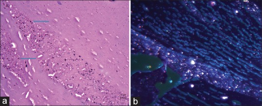
Figure 3.

H and E (Hematoxylin and eosin) sections studied from the lens nucleus tissue (a) shows diffuse deposition of black granules (pointed by blue colored arrows) suggestive of silver particles which are refractable in dark field illumination (b)
