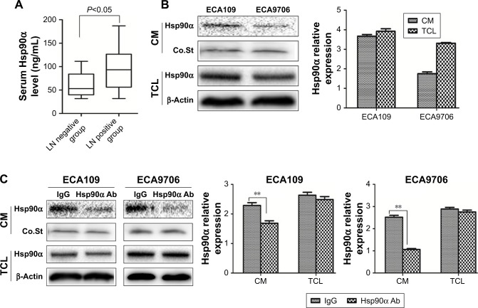
Figure 1.
The serum Hsp90α level of ESCC patients and secretion of Hsp90α from ESCC cells.
Notes: (A) The serum Hsp90α level before treatment in positive LN group was much higher (P<0.05) than that of LN negative group. (B) ECA109 and ECA9706 were plated in 6 cm dishes. The media were replaced with serum-free medium when cells grew to 80% confluence. After 8 hours, the CM were collected and concentrated 10-fold. The amounts of Hsp90α in CM and TCL were analyzed by Western blotting. Coomassie blue staining as a loading control for CM and β-actin was used as loading control for TCL. (C) Control IgG (10 µg/mL) or Hsp90α Ab (10 µg/mL) was added to the serum-free medium of ESCC cells. After 24 hours of treatment, the amount of Hsp90α in CM and TCL was analyzed. Data shown as mean ± SD, **P<0.001.
Abbreviations: CM, conditioned media; Co.St, Coomassie blue staining; ESCC, esophageal squamous cell carcinoma; Hsp90α Ab, Hsp90α neutralizing antibody; LN, lymph node; TCL, total cell lysate.

