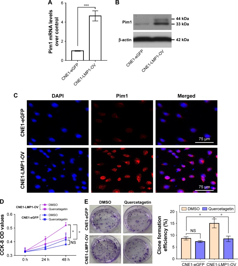
Figure 4.
LMP1-induced Pim1 expression promotes NPC cell proliferation.
Notes: (A) Enhanced Pim1 mRNA levels in LMP1 stably expressing cells. ***P<0.001. The experiment was repeated three times (n=3). (B) LMP1 mainly upregulates the 33 kDa Pim1 protein but not the 44 kDa protein in NPC cells. The experiment was repeated three times (n=3) and the representative bands are shown. (C) Expression and cellular location of Pim1 protein in NPC cells. Antigens for Pim1 were colorized with TRITC-coupled IgGs. Bars, 75 µm. (D) Cell viability of LMP1-overexpressing cells and control cells treated with Pim1 inhibitor quercetagetin, assessed by CCK-8 assay. *P<0.05. Each group was repeated in six wells (n=6). (E) Clone formation efficiency of quercetagetin-treated cells. *P<0.05. Each group was repeated in three wells (n=3).
Abbreviations: CCK-8, cell counting kit-8; DAPI, 4′,6-diamidino-2-phenylindole; DMSO, dimethyl sulfoxide; NS, no significance; TRITC, tetramethylrhodamine isothiocyanate.

