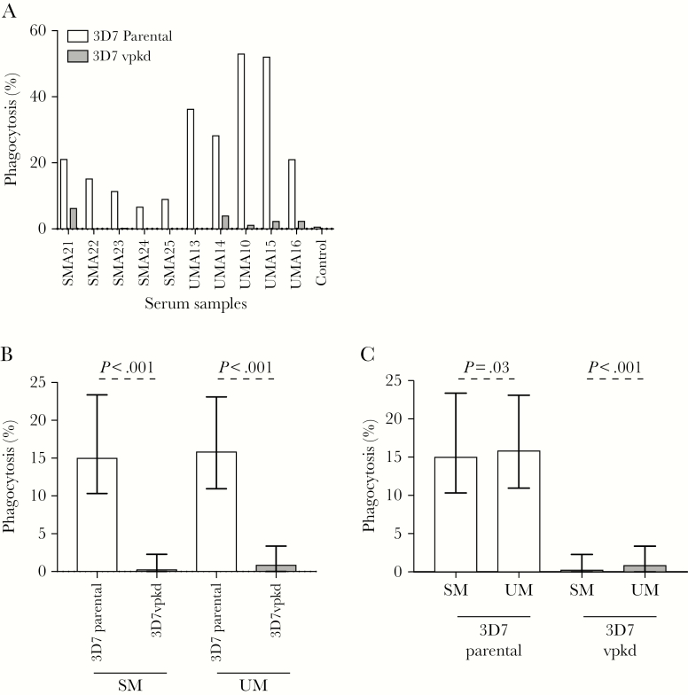
Figure 5.
Opsonic phagocytosis of Plasmodium falciparum–infected erythrocytes (IEs) by undifferentiated THP-1 monocytes. A, Representative selection of serum samples collected during acute infection was tested for opsonic phagocytosis activity to 3D7parental and 3D7vpkd parasites. Assay was performed once (n = 235 for severe malaria [SM]; n = 213 for uncomplicated malaria [UM]); bars represent the mean level of phagocytosis as a percentage of positive control. B, Opsonic phagocytosis activity of serum antibodies was markedly reduced with 3D7vpkd parasites compared to 3D7parental, for SM and UM. Bars represent the median and interquartile range (IQR) of samples that were classified as antibody positive to 3D7parental (n = 80/235 for SM; n = 96/213 for UM); P values were calculated using a paired Wilcoxon signed-rank test. C, There was a higher level of opsonic phagocytosis activity with samples from UM compared to SM. Bars represent the median and IQR of samples that were classified as opsonic phagocytosis activity that is positive to 3D7parental (n = 80/235 for SM; n = 96/213 for UM); P value was calculated using an unpaired Mann–Whitney test. Abbreviations: SM, severe malaria; UM, uncomplicated malaria.

