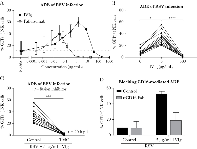
Figure 2.
Antibody-dependent enhancement of respiratory syncytial virus (RSV) infection in natural killer (NK) cells. A, Infection of adult primary NK cells in the presence of RSV-X-GFP7-antibody complexes (with the indicated concentration of intravenous immunoglobulin [IVIg] or palivizumab). Graph depicts the geometric mean and standard deviation (SD) of 3 donors from 1 experiment. Dashed line depicts the geometric mean percentage green fluorescent protein (GFP)–positive cells in absence of antibodies (no antibodies). B, Infection of NK cells with RSV-X-GFP7 or (sub)neutralizing RSV-antibody complexes (5 µg/mL or 500 µg/mL IVIg). Data were pooled from 6 independent experiments and each set of paired data points represents an individual donor (n = 14). C, Infection of NK cells with subneutralizing RSV-antibody complexes (5 µg/mL IVIg) in the absence or presence of a fusion inhibitor (TMC). Data were pooled from 6 independent experiments and each set of paired data points represents an individual donor (n = 14). D, Infection of NK cells in the absence or presence of subneutralizing RSV-antibody complexes (5 µg/mL IVIg) with 50 µg/mL CD16-blocking Fab fragments. Graph depicts the geometric mean and SD of 3 donors from 1 experiment. All GFP measurements were performed by flow cytometry at 20 hours postinfection. Nonparametric Friedman test with Dunn multiple comparisons test was used for comparisons between multiple conditions (*P < .05, ****P < .0001). Wilcoxon signed-rank test was used for comparison between 2 conditions (***P < .001). Abbreviations: Abs, antibodies; ADE, antibody-dependent enhancement; GFP, green fluorescent protein; h.p.i., hours postinfection; IVIg, intravenous immunoglobulin; NK, natural killer; RSV, respiratory syncytial virus.

