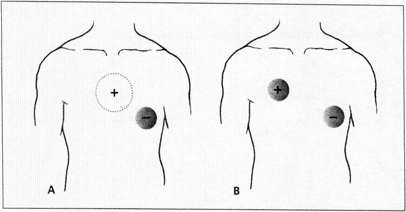
Figure 1.
Electrode positions in transcutaneous pacing are shown here. Most commonly, the negative electrode is placed on the anterior chest wall, centered on the apex, or at the lead V3 position along the left sternal border. The positive electrode is placed on the patient’s back between the spine and left scapula, directly posterior to the anterior electrode (A). In some patients, an alternative is to place the positive electrode on the right side of the chest at the lead V1 position and the negative electrode on the cardiac apex (B).

