Fig. 4.
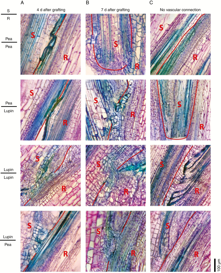
Vascular development in heterografts 4 and 7 d after grafting. Photographs of 30 μm thick longitudinal sections of the graft junction in reciprocal grafts between lupin and pea scions (S) and rootstocks (R). The graft junctions were harvested (A) 4 d or (B) 7 d after grafting. Wedge grafts were carried out with 1- to 2-week-old seedlings as described by Foo et al. (2015). The pea scions and rootstocks were severed in the first or second internode, lupin rootsocks in the epicotyl, and lupin scions in the hypocotyl. Sections were fixed in 4 % paraformaldehyde and embedded in polyethylene glycol (PEG) 1000 (using a method based on Paciorek et al., 2006) prior to sectioning and staining with toluidine blue and safranin O. The red line shows the junction between the scion and stock tissue. (C) Examples of grafts showing regions of no vascular reconnection for comparison.

