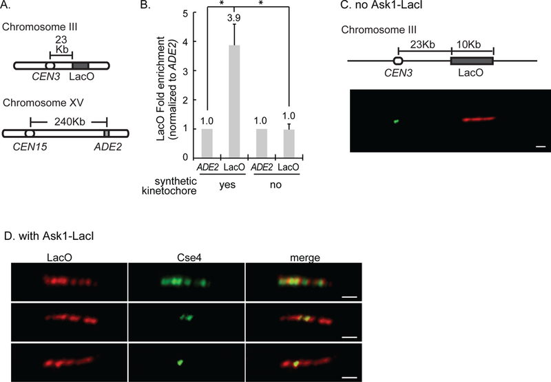
Figure 1. Cse4 localizes to the site of synthetic kinetochore assembly.

A) The three loci analyzed by ChIP. B) Quantification of ChIP-qPCR averaged from three experiments ± S.D, showing the LacO enrichment over non-specific binding at the ADE2 locus. * represents statistical significance. C, D) Representative images of chromatin fiber immunofluorescence. Strains have the LacO array and express GFP-Cse4 and mCherry-LacI. Cse4 and LacI were detected using anti-GFP and anti-mCherry antibodies. Scale bar 1μm. C) Chromatin fiber from a strain without Ask1-LacI showing Cse4 at CEN3 and LacO ~23Kb away. D) Images of chromatin fibers from a strain with Ask1-LacI with Cse4 along the entire array (top), part of the array (middle), and in one focus (bottom).
