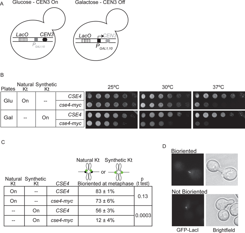
Figure 2. Cse4-Myc disrupts synthetic kinetochore function.

A) Depiction of natural kinetochore function in cells with the GAL1,10 promoter at CEN3 (PGAL1,10CEN3) and grown in glucose versus galactose-containing medium. B) Serial dilutions of cultures spotted on glucose and galactose plates at 25, 30, and 37°C. C) The percent biorientation of natural or synthetic kinetochores in metaphase-arrested cells grown at 37°C (average of 3 experiments ± S.D., 100 cells counted per experiment). D) Images show a cse4-Myc cell with (top) and without (bottom) bioriented synthetic kinetochores.
