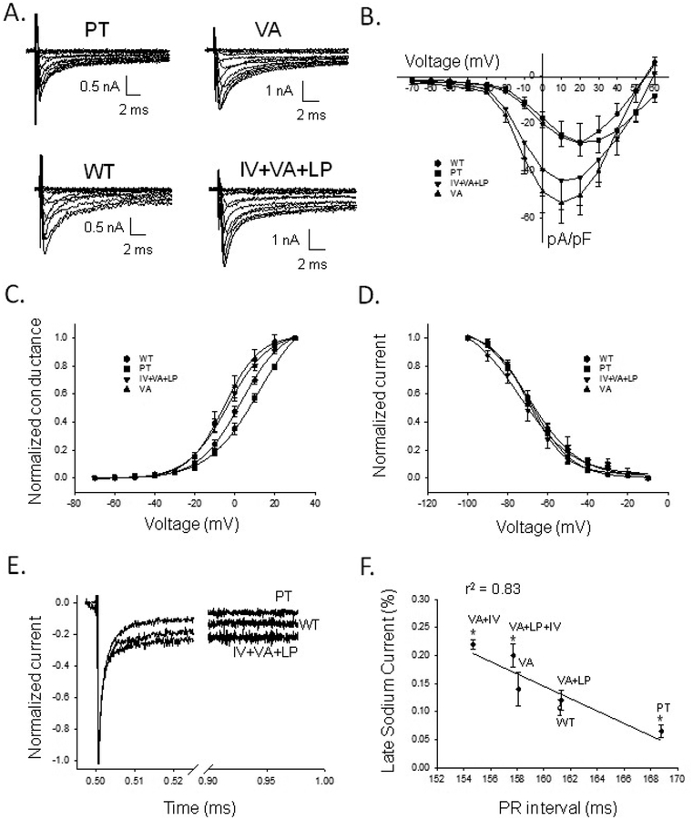
Figure 2. Electrophysiological characteristics of Nav1.8 channel haplotypes.
A) Four haplotype Nav1.8 whole-cell sodium currents recorded from N2a cells in the presence of 150 nM tetrodotoxin. Currents were elicited from a holding potential of −100 mV with depolarizing steps between −70 and +60mV in 10mV increments. B) Current density (pA/pF) versus voltage for WT, PT, VA, and IV+VA+LP constructs. C) Conductance versus voltage for WT, PT, VA, and IV+VA+LP, fit with a Boltzmann function (see Supplemental Methods). V1/2 for PT was shifted rightward, and VA and IV+VA+LP were both shifted leftward compared to WT. Slope factors (k) were not significantly different. D) Voltage-dependent inactivation as normalized current versus voltage. The V1/2 and k for the haplotype variants were not significantly different from WT. E) Normalized INa,L of PT, WT, and IV+VA+LP, measured from a holding current of −100mV with pulse to +20mV for 475ms. PT shows a smaller and VA+LP+IV a greater INa,L compared to WT, respectively. F) Percent INa,L versus PR interval. The percent INa,L decreases moving from haplotypes with short to long PR intervals. The data were significantly correlated by the fitted line (r2=0.83, P=0.01). *: statistically different from WT (P>0.05).

