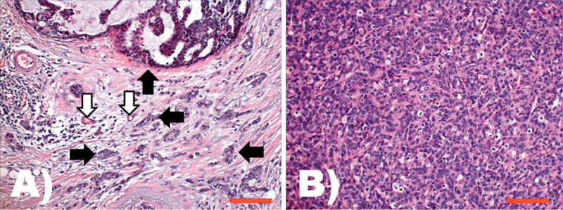
Figure 1.

Comparison of phenotypic features of cancer cells in their natural environment versus a xenograph implanted into a mouse. Both A and B are images taken from H&E stained slides at the same original magnification (200 ×); the scale bars represents 200 µm. A: Typical aspect of a ductal carcinoma, the most common histological type of human breast cancer. Tumor epithelial cells are arranged in diverse architectural patterns (e.g. see upper side of this field and black arrows). The tumor epithelial cells are in intimate relation with the stroma that is composed of multiple non-tumor cell types including blood vessels (white arrows), fibroblasts, and inflammatory cells. The functional unit of tumor and cancer associated mesenchymal cells can be considered as the cancer histion. B: Morphological phenotype of human breast ductal carcinoma isolated and cultured as a cell line, and subsequently implanted as a xenograft into a mouse. The morphology of this model system is distinctly different from that observed in patients; here the ductal carcinoma cells grow as solid sheets, showing higher atypia, increased mitotic activity, and a different shape and size than in their natural tissue environment. The functional structure of the tumor microenvironment – the cancer histion – is lost in this model.
