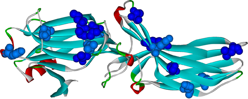
Fig. 4. Elements of arrestin-1 that move upon rhodopsin binding.
Bovine arrestin-1 structure in it basal conformation (PDB ID: 1CF1 (Hirsch et al., 1999)) is shown, with the elements that move upon rhodopsin binding (Kim et al., 2012) colored dark red. These include the finger loop (residues 67–78 (Hanson et al., 2006)), the 139-loop (Kim et al., 2012) (a.k.a. the middle loop in arrestin-2 (Shukla et al., 2013)), as well as loops at the distal tips of the N-domain (residues 155–168) and C-domain (residues 336–344).

