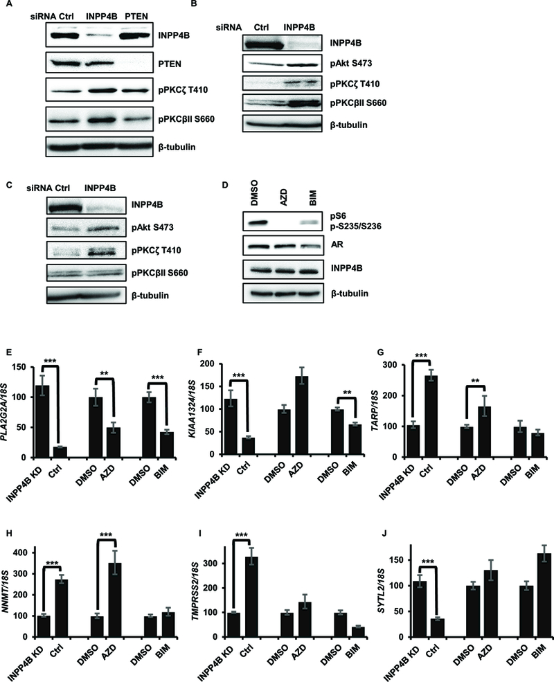
Figure 4. Suppression of Akt and PKC signaling contributes to INPP4B regulation of AR transcriptional activity.

(A) VCaP cells were transfected with control, INPP4B, or PTEN specific siRNAs. Expression levels of INPP4B, PTEN, pPKCζ, pPKCβII and β-tubulin were measured by Western blotting. (B) LNCaP cells were transfected with control or INPP4B siRNAs for 48 hours in medium supplemented with 10% FBS. Cellular lysates were assayed for INPP4B, pAkt, pPKCζ, pPKCβII and β-tubulin by Western blotting. (C) C4–2 cells were transfected and assayed in parallel with B. (D) LNCaP cells were plated in complete medium and treated with indicated inhibitors for 8 hours. Protein extracts were assayed for AR, INPP4B, pS6, and tubulin levels by Western blotting. (E-J) LNCaP cells were plated in medium supplemented with 10% CSS with either vehicle or 1 nM R1881. Twenty four hours later cells were treated with vehicle (DMSO), 5μM AZD5363, or 2 μM BIM-I for an additional 8 hours. In parallel, LNCaP cells were transfected with control or INPP4B siRNA for 48 hours in 10% CSS medium supplemented with 1 nM R1881. RNA was purified, reverse transcribed, and expression levels of AR regulated genes PLA2G2A (E), KIAA1324 (F), TARP (G), NNMT (H), TMPRSS2 (I) and SYTL2 (J) compared by RT-qPCR. ** represents p < 0.01; *** represents p < 0.001.
