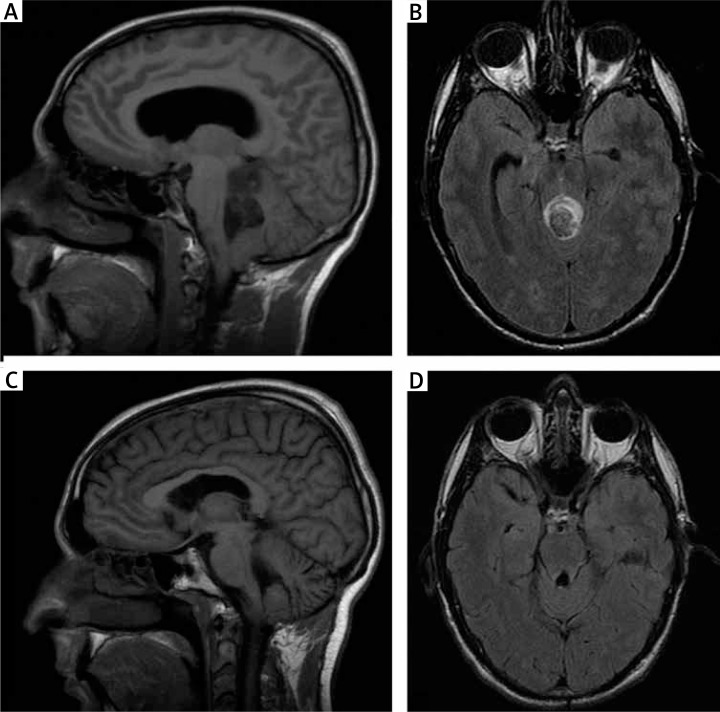
Fig. 1.
A–B) Preoperative MRI. T1-weighted sagittal and T2-weighted axial images demonstrating the tumour mass with a cystic component and extension into the floor of the fourth ventricle and to the supravermian cistern. Partial obstruction of the fourth ventricle and secondary obstructive hydrocephalus is also observed. C–D) Two-year postoperative MRI. No apparent residual tumour is shown at T1-weighted sagittal and T2-weighted axial images

