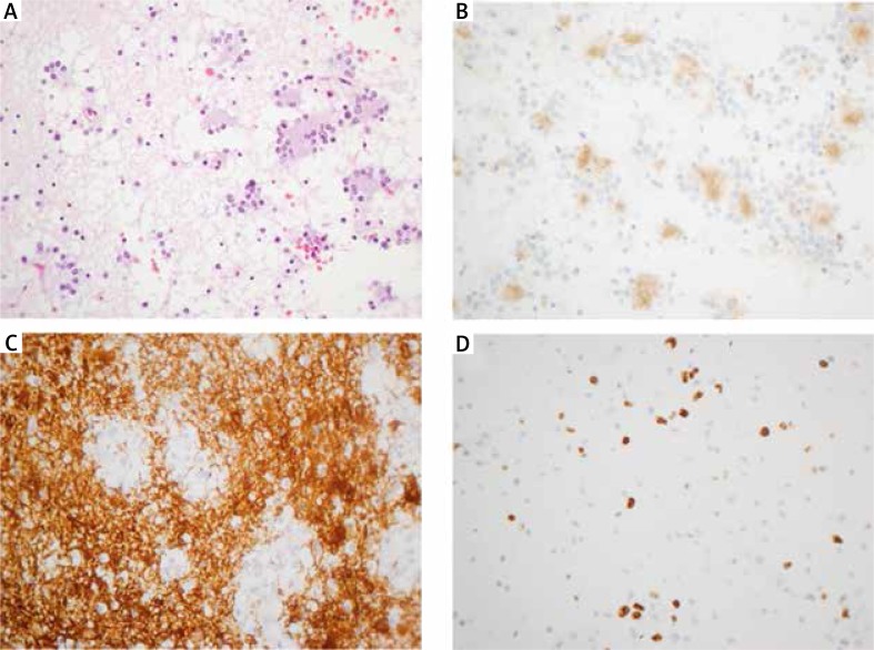
Fig. 2.
Microscopic features of the rosette-forming glioneuronal tumour. A) Histological features of the tumour showing both a neurocytic and an astrocytic component. Neurocytic rosettes are formed by the neurocytic components. B) The eosinophilic core at the centre of the neurocytic rosettes displays strong positive staining with synaptophysin. C) The astrocytic components of the tumour showed that the tumour cells had bipolar and spindle processes with positive immunostaining of GFAP. D) The MIB-1 labelling index was about 5–7%. Original magnifications 400×

