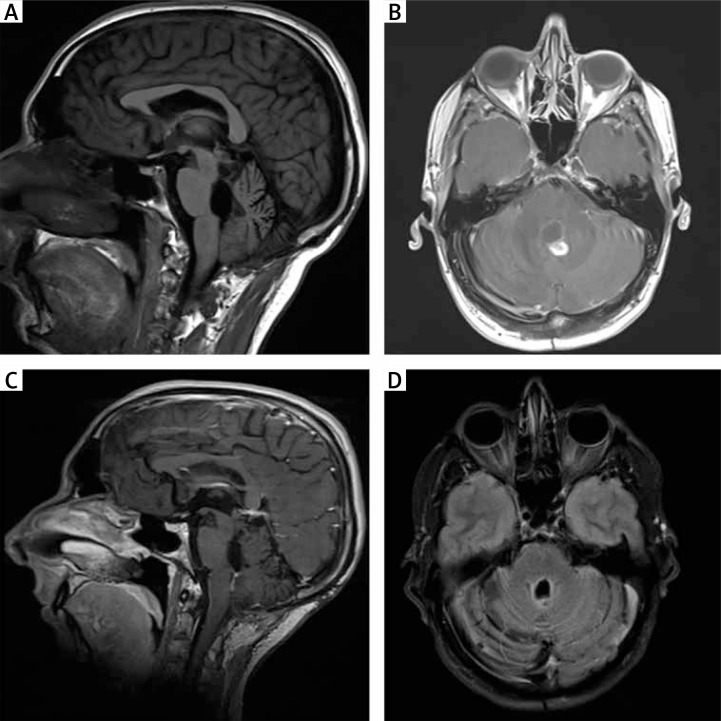
Fig. 3.
A–B) MRI scans 48 months after the initial procedure. T1-weighted sagittal and T1-weighted axial contrast enhanced images reveal a nodular lesion close to the roof of the fourth ventricle. C–D) T1-weighted sagittal and T2-weighted axial MRI images two years after radiosurgery show stabilisation of the lesions

