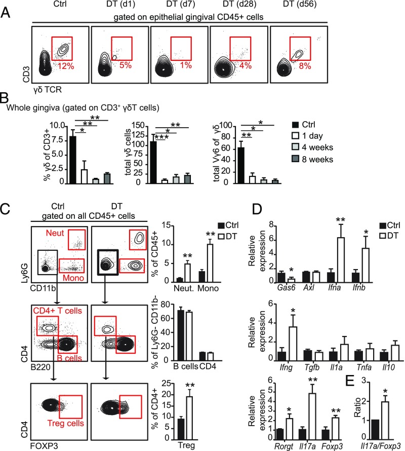
Fig. 6.
Prolonged ablation of γδT cells increases gingival inflammation. (A) Repopulation kinetics of γδT cells in the gingiva and ear skin following single administration of DT to Tcrd-GDL mice. FACS plots demonstrate the frequencies of epithelial γδT cells; at least three mice were analyzed at each time point. (B) Bar graphs represent the mean + SEM of pooled data from three independent experiments (n = 4–10 mice per experiment) of frequencies of γδT cells among CD3 as well as total numbers of γδT cells and Vγ6+ γδT cells in the gingiva. Three to 10 mice were analyzed at each time point. (C and D) Adult Tcrd-GDL mice were injected intraperitoneally with DT on a weekly basis for 5 mo; gingival tissues were then collected and analyzed. (C) Representative FACS plots and graphs demonstrate the alterations in the frequencies of various leukocytes subsets in the gingiva due to the absence of γδT cells. (D) Relative expression of the indicated genes was quantified by qRT-PCR. Graphs present the transcript levels normalized to control group (Ctrl) depicted as the mean values + SEM (n = 5–6). (E) Il17a/Foxp3 expression level ratio in DT-treated and control groups presented as the mean values + SEM (n = 5). Representative data of two independent experiments are presented. *P < 0.05, **P < 0.01, ***P < 0.001.

