Fig. 5.
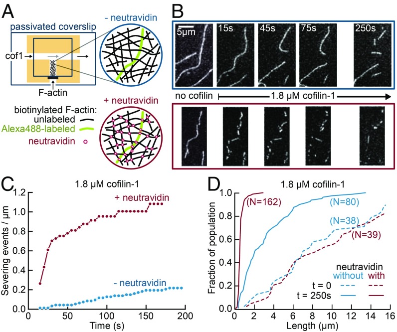
Enhanced severing of interconnected actin filament networks. (A) Experimental setup, using T-shaped chambers, seen from above. A solution of biotinylated F-actin, containing one fluorescently labeled filament for 20 unlabeled filaments, was first injected in the short channel. The filaments could either be left nonconnected (Top, blue) or be cross-linked by injecting neutravidin through the main channel (Bottom, red). Cofilin-1 (unlabeled) was then injected in the main channel and F-actin severing was observed in the same region 500 µm away from the channel junction. All solutions were supplemented with 0.15% methylcellulose to maintain filaments close to the surface, and 0.25% BSA to maintain a good surface passivation. (B) Time-lapse of individual Alexa-488–labeled filaments, within a meshwork of unlabeled filaments, before and after filling the main channel with 1.8 µM cofilin (at time t = 0). Filaments are either nonconnected (Top, blue) or cross-linked by neutravidin (Bottom, red). (C). Quantification of severing events, cumulated over time, for nonconnected (blue, 10 filaments with a total initial length of 102 µm) and interconnected (red, five filaments with a total initial length of 60 µm) filaments, following the introduction of 1.8 µM cofilin in the main channel. (D) Cumulative length distributions (i.e., showing the fraction of a filament population having a length smaller than a given value) before exposure to cofilin (dashed lines) and 250 s after flowing 1.8 µM cofilin in the main channel (solid lines), for nonconnected (blue, initial population of 38 filaments, final population of 80 observable filament fragments) and interconnected filaments (red, initial population of 39 filaments, final population of 162 observable filament fragments).

