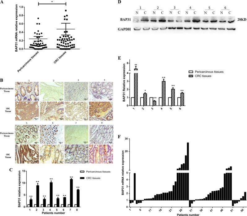
Fig. 1. BAP31 expression was significantly elevated in CRC tissues compared to pericarcinous tissues.
a The relative expression of BAP31 mRNA in CRC tissues of 57 cases compared to pericarcinous tissues as detected by real-time PCR (*P < 0.05). b, c The relative expression of BAP31 protein in CRC tissues compared to pericarcinous tissues as detected by immunohistochemistry (**P < 0.01; 1–6: six pairs of resected CRC specimens were randomly selected from stage II and stage III patients; samples 1–3 were in stage II, and samples 4–6 in stage III) and western blotting (**P < 0.01; N, pericarcinous tissues; C, colorectal cancer; 1–4: eight pairs of clinically resected CRC specimens randomly selected from stage II and stage III patients; samples 1–4 were in stage II, and samples 5–8 in stage III). d, e The relative expression of BAP31 protein in CRC tissues compared to pericarcinous tissues as detected by immunohistochemistry (scale bar, 50 μm; **P < 0.01). (f) The expression of BAP31 in CRC tissues of patients with different clinic stages: 1–3, stage I; 4–29, stage II; 30–50, stage III; 51–57, stage IV. CRC, colorectal cancer

