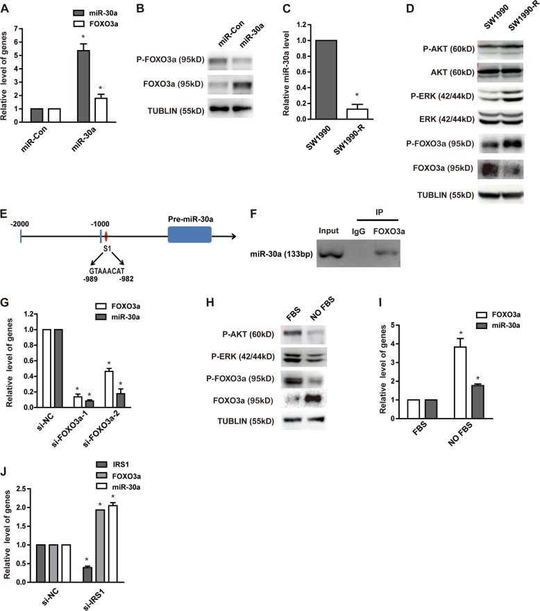
Fig. 6. miR-30a and FOXO3a positively regulate each other to synergistically work as tumor suppressor.
a, b SW1990 cells were transfected with indicated constructs. FOXO3a expression in SW1990 cells with or without miR-30a overexpression was detected by qRT-PCR (a). *P < 0.05, compared with control conditions. Protein expression of phospho-FOXO3a and total FOXO3a level were detected by western blot (b). c MiR-30a expression in SW1990-R cells and its parental cells was detected by qRT-PCR. *P < 0.05, compared with SW1990. d Gemcitabine-induced changes in protein expression between SW1990 cells and its parental cells were examined by western blot. e Computational prediction of FOXO3a binding site (S1) on 5’ miR-30a regulatory region. f Representative anti-FoxO3a ChIP-PCR showing FoxO3a binding to the miR-30a promoter. g Knockdown of FOXO3a with two different FOXO3a siRNAs (si-FOXO3a-1, si-FOXO3a-2) in SW1990 cells. MiR-30a level was determined by qRT-PCR subsequently. *P < 0.05, compared with control conditions. h, i SW1990 cells were treated with or without 10% FBS after overnight serum starvation. The P-AKT, P-ERK, P-FOXO3a, and FOXO3a in cell lysates were tested by western blot (h); mRNA level of FOXO3a and miR-30a were detected by qRT-PCR (i). *P < 0.05, compared with FBS group. j Transient knockdown of IRS1 with siRNA in SW1990 cells. MiR-30a and FOXO3a level were detected by qRT-PCR subsequently. *P < 0.05, compared with control conditions. Points, mean values for three independent experiments; error bars, ±SEM

