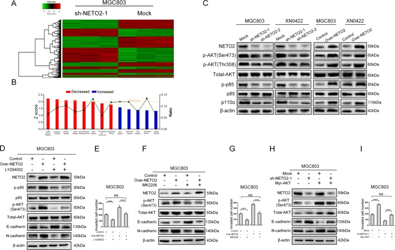
Fig. 3. NETO2 activates PI3K/AKT pathway to induce EMT in GC cells.
a Microarray heatmap showed differentially expressed genes in mock and sh-NETO2–1 cells. The color key for the normalized expression data was shown at the top of the microarray heatmap (green means downregulated genes and red represents upregulated genes). b Ingenuity Pathway Analysis (IPA) of the most significantly changed classical pathways after knockdown of NETO2 in gastric cancer cells. All signaling pathways were sorted according to the z score (left y-axis). The ratio represented the relative number of differentially expressed genes in a specified signaling pathway (right y-axis). c Western blotting analysis showed reduced phosphorylation levels of AKT and p85 in sh-NETO2–1 cells, while increased phosphorylation levels of AKT and p85 in Over-NETO2 cells compared to their control cells, respectively. d Western blotting analysis showed that treatment with LY294002 (10 μM) attenuated NETO2-enhanced phosphorylation of AKT and inhibited NETO2-induced EMT in Over-NETO2 cells. e Quantification of transwell invasion assay showed that treatment with LY294002 (10 μM) attenuated the enhanced invasive ability in Over-NETO2 cells. f Western blotting analysis showed that treatment with MK2206 (5 μM) attenuated NETO2-enhanced phosphorylation of AKT and inhibited NETO2-induced EMT in Over-NETO2 cells. g Quantification of transwell invasion assay showed that treatment with MK2206 (5 μM) attenuated the invasive ability in Over-NETO2 cells. h Western blotting analysis showed that transfection with myristoylated (myr) -AKT increased the phosphorylation level of AKT and induced EMT in NETO2-knockdown cells. i Quantification of transwell invasion assay showed that transfection with myr-AKT increased the invasive ability in NETO2-knockdown cells. ***p<0.001; NS no significant

