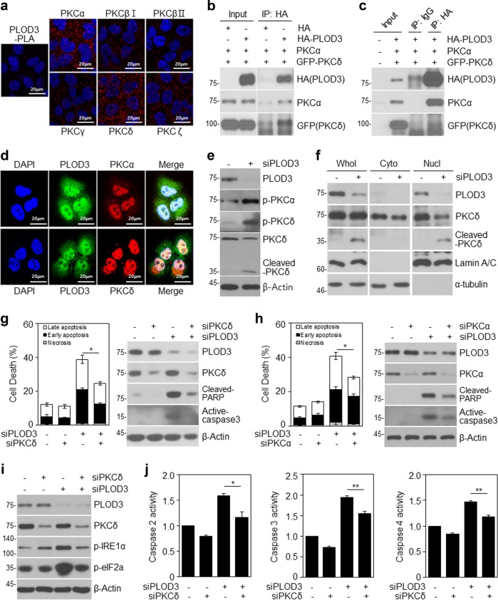
Fig. 5. PLOD3 knockdown induces activation of PKCs and PLOD3 directly interacts with PKCs.
a R-H460 cells were fixed and incubated with a mouse anti-PLOD3 antibody together with rabbit antibodies against PKCs, followed by in situ proximity ligation assay analysis. Representative confocal images of cells with proximity ligation assay-positive signals (red dots). b, c R-H460 cells were transfected with HA-PLOD3, PKCα, or GFP-PKCδ. Cell lysates were immunoprecipitated with normal IgG (a negative control) or HA antibody, and then immunocomplexes were resolved by SDS-PAGE and immunoblotted with antibodies against HA, PKCα, and GFP. d Immunostaining analysis of the localization of PKCs in R-H460 cells. Cells were fixed with 4% paraformaldehyde and immunostained for PLOD3 (green), PKCα (red), and PKCδ (red). DNA was visualized by DAPI staining. e PLOD3 or control siRNA was transfected into R-H460 cells; 24-h post-transfection, cell lysates were prepared and used in immunoblotting with antibodies targeting PLOD3, p-PKCα, PKCδ, p-PKCδ, and β-actin. f Subcellular fraction analysis of R-H460 cells following siCON or siPLOD3 treatment. Proteins in each fraction were resolved by SDS-PAGE and immunoblotted with antibodies against PLOD3, PKCδ, lamin A/C, and α-tubulin. g R-H460 cells were transfected with 40 nM siCON or siPLOD3 and/or siPKCδ for 48 h. Cell death in R-H460 cells was determined by AV/PI staining (left). Protein levels of the indicated proteins were determined by western blotting (right). *P < 0.05. h R-H460 cells were transfected with 40 nM siCON or siPLOD3 and/or siPKCα for 48 h. Cell death in R-H460 cells was determined by AV/PI staining (left). Protein levels of the indicated proteins were determined by western blotting (right). *P < 0.05. i Protein levels of PLOD3, PKCδ, p-eIF2α, and p-IRE1 α were determined by western blotting. j Caspase-2, caspase-3, and caspase-4 activities in R-H460 cells (treated as in b) were determined by caspase activity assay. Data were collected using the Multiskan EX at 405 nm. *P < 0.05; **P < 0.01

