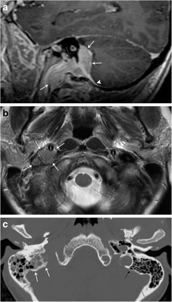
Fig. 10.

Meningioma. a Sagittal T1-weighted post-contrast MR image demonstrates an enhancing mass (white arrows) extending from the posterior fossa through the jugular foramen (black arrows) into the right carotid space. An enhancing dural tail is seen (arrowhead). b Axial T2-weighted MR image demonstrates the mass to be intermediate in signal (white arrows), anteriorly displacing the internal carotid artery (i). The normal position of the left internal carotid artery is also seen. c Axial non-contrast CT image demonstrates hyperostosis of the bone adjacent to the known lesion (white arrows). Note the thickness of the normal left mastoid bone cortex (black arrow)
