
Fig. 2.
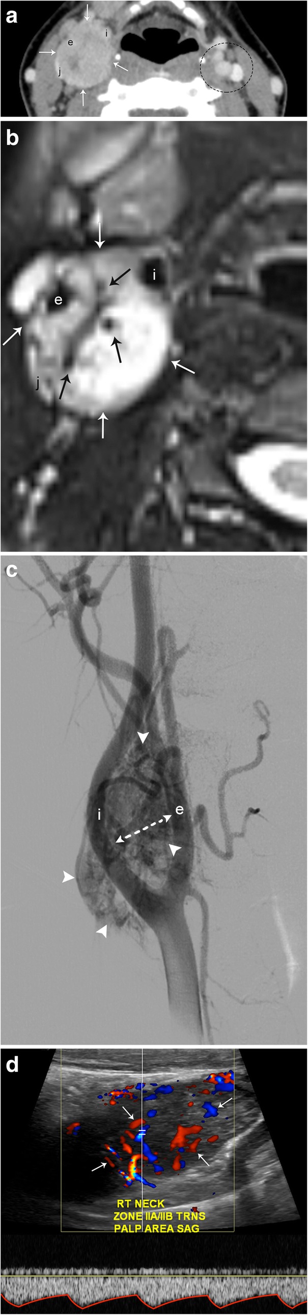
Carotid body tumor. a Axial CT image demonstrates a well-circumscribed, enhancing mass centered in the right carotid space (arrows) that causes splaying of the right internal (i) and external (e) carotid arteries. There is mass effect on the internal jugular vein (j). The left carotid space (dotted circle) demonstrates the normal relationship of the carotid and jugular vessels. b Axial T2-weighted MR image shows a well-circumscribed hyperintense mass (white arrows) with multiple internal flow voids (black arrows). c DSA demonstrates an intensely hypervascular mass (arrowheads) centered at the carotid bifurcation. Again seen is splaying of the internal and external carotid arteries. d Spectral Doppler ultrasound confirms hypervascularity (arrows) and internal arterial flow (spectral tracing) of this lesion