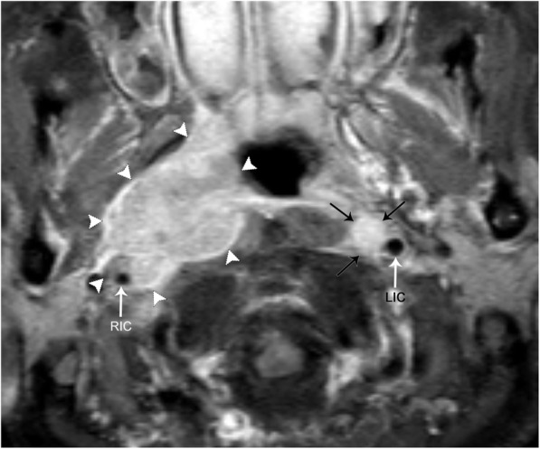
Fig. 22.

Direct spread of non-HPV-associated squamous cell carcinoma into the carotid space. Axial contrast-enhanced, fat-suppressed T1 image demonstrates a large, avidly enhancing and infiltrating mass that invades the right carotid space (white arrowheads). The right carotid artery (RIC) is encased, narrowed, and laterally displaced when compared to the left internal carotid artery (LIC). Encasement of the carotid artery upstages the disease to T4b. There is also a left retropharyngeal lymph node (black arrows), just medial to the LIC
