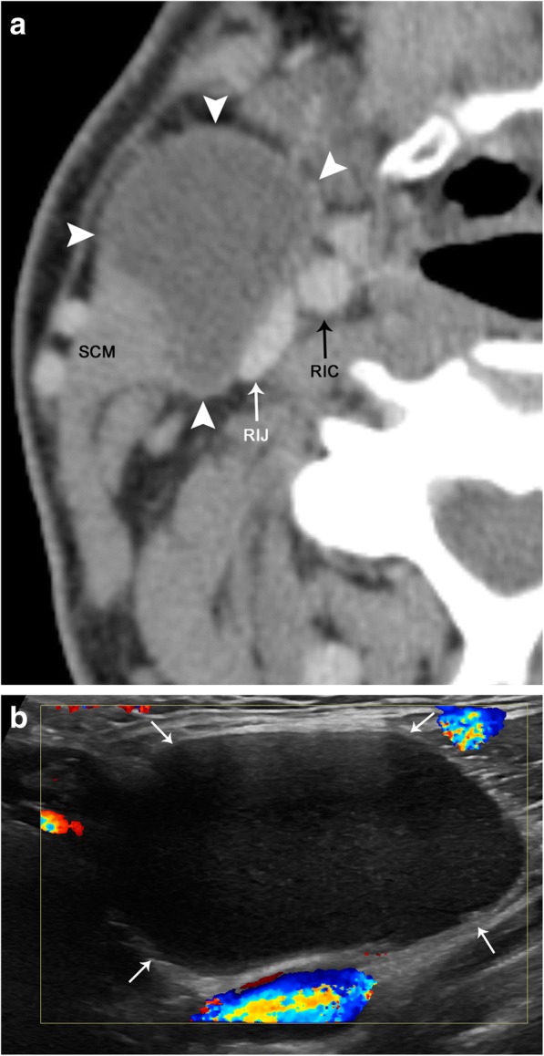
Fig. 23.

Second branchial cleft cyst. a Axial contrast-enhanced CT image demonstrates a well-circumscribed and hypoattenuating lesion (arrowheads) along the anterior margin of the right sternocleidomastoid muscle (SCM) and anterolateral to the right carotid space, internal carotid artery (RIC), and internal jugular vein (RIJ). b Color Doppler ultrasound image demonstrates that this lesion (arrows) is hypoechoic with low-level echoes. There is no internal vascularity or peripheral hyperemia
