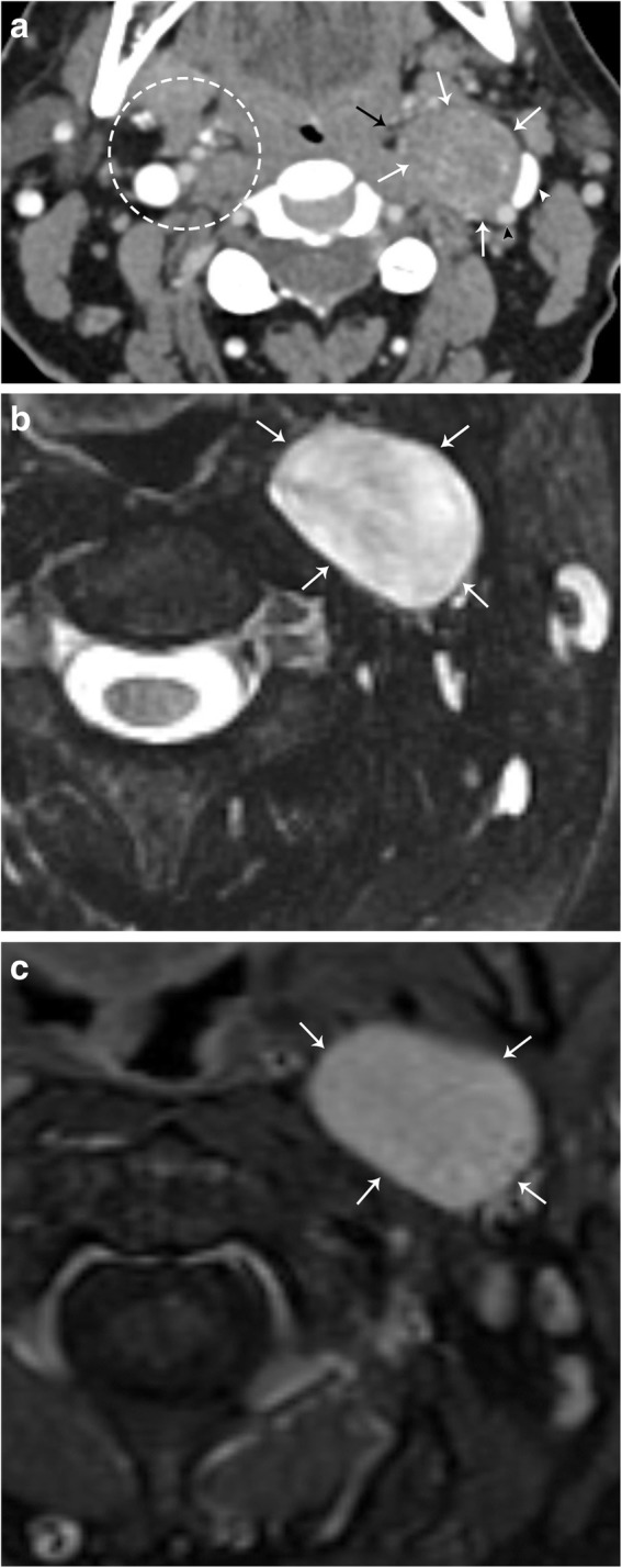
Fig. 9.

Sympathetic plexus schwannoma. a Axial CT image demonstrates a well-circumscribed mass (white arrows) that laterally displaces the left internal carotid artery (black arrowhead) and left internal jugular vein (white arrowhead). The left parapharyngeal fat is medially displaced (black arrow). Note the normal configuration of the right carotid space and parapharyngeal fat (dotted circle). b Axial T2 fat-suppressed MR image demonstrates homogenous hyperintensity of this mass (white arrows). c Axial T1 post-contrast fat-saturated MR image demonstrates homogenous enhancement of the lesion. Note the anteromedial location of the lesion relative to the carotid vessels, indicating sympathetic plexus origin
