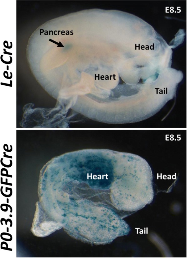
Fig. 5.

P0-3.9GFPCre E8.5 embryos exhibit extensive CRE-mediated recombination prior to lens placode formation. At E8.5, X-Gal staining in Le-Cre embryos (top) remained restricted to the developing pancreas. In contrast, P0-3.9GFPCre embryos (bottom) exhibited many patches of X-Gal stained tissue throughout the embryo, with particularly high numbers of blue clones in the developing heart region
