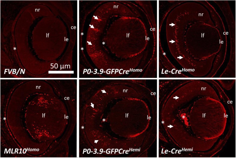
Fig. 6.

Immunohistochemical detection of CRE protein in P0-3.9GFPCre, Le-Cre, and MLR10 transgenic mice at E15.5. MLR10 exhibited CRE protein expression specifically in the nuclei of differentiating lens fiber cells (lf) within the eye at E15.5, while P0-3.9GFPCre, Le-Cre lenses showed obvious CRE protein in the epithelium of both the lens (le) and cornea (ce) and numerous cells within the developing neural retina (nr) indicated by arrows. Notice that the homozygous Le-Cre (Le-CreHomo) lens is specifically small and misshapen relative to the lenses from the other genotypes. The FVB/N lens exhibits no specific staining with the anti-CRE antibody and serves as a negative control. Autofluorescence in blood cells in the choroid and tunica vasculosa lentis represent non-specific signal (asterisks). Transgenic homozygotes and hemizygotes are indicated by homo and hemi, respectively
