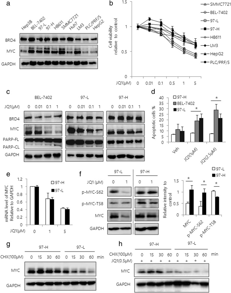
Fig. 2.
JQ1 selectively inhibits HCC cell tumor growth. a Western blot analysis of BRD4 and MYC expression in 10 HCC cell lines. GAPDH was used as a loading control. b Cell viability curves are shown for serial dilutions of JQ1 in 8 HCC cell lines. Cell viability was determined at 48 h after treatment using Cell Titer-Glo. c Western blot analysis of BEL-7402, 97-L and 97-H cells treated with serial dilutions of JQ1 for 48 h. Total lysates were subjected to the indicated antibodies. d BEL-7402, 97-L and 97-H cells were treated with varying dose of JQ1 for 48 h. Apoptosis was assessed by Annexin V / PI double staining. Quantification of apoptotic cells was determined based on Annexin V positive cells. e 97-L and 97-H cells were treated with JQ1 for 48 h. MYC mRNA expression level was analyzed by qRT-PCR. f Western blot analysis of 97-L and 97-H cells treated with 1 μM JQ1 for 48 h. Expressions of MYC, p-MYC-Ser62 and p-MYC-Thr58 were examined. Band intensities were quantified by Image J software and graphed at the right side. GAPDH was used as a loading control. Western blot analysis of 97-L and 97-H cells incubated with 100 μM CHX in the absence (g) or presence (h) of JQ1. Data are presented as mean ± s.d. *p < 0.05

