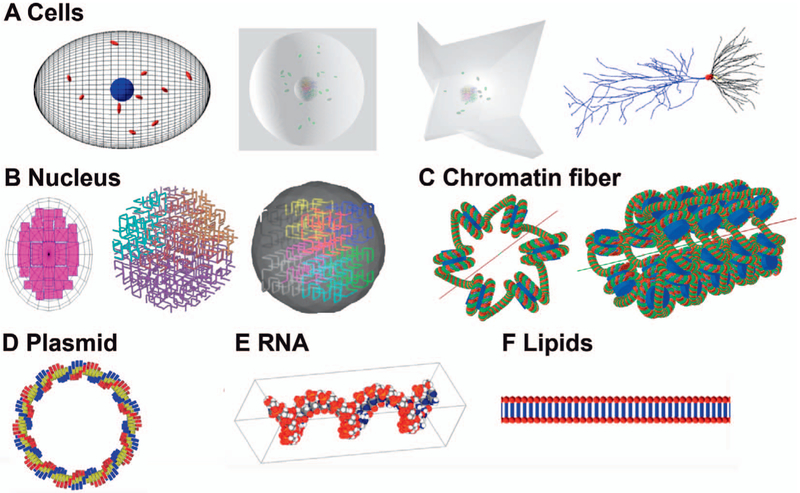
FIG. 4.
Geometries available in TOPAS-nBio. Panel A: An ellipsoid cell shown with a nucleus (blue) and mitochondria (red), a spherical and a fibroblast cell with nucleus and mitochondria. Also shown is a hippocampal neuron with a soma (red) and dendrites (black and blue). Panel B: Three full nucleus models, one based on the Geant4-DNA example (left side) and two different fractal models (center and right side). Panel C: A chromatin fiber consisting of nucleosomes each composed of histone proteins (blue) wrapped by two turns of a double helix DNA (green and red). Panel D: A circular plasmid consisting of 100 basepairs. Panel E: RNA strand recreated using the TOPAS-nBio interface to the protein database. Panel F: A lipid (membrane) layer.

