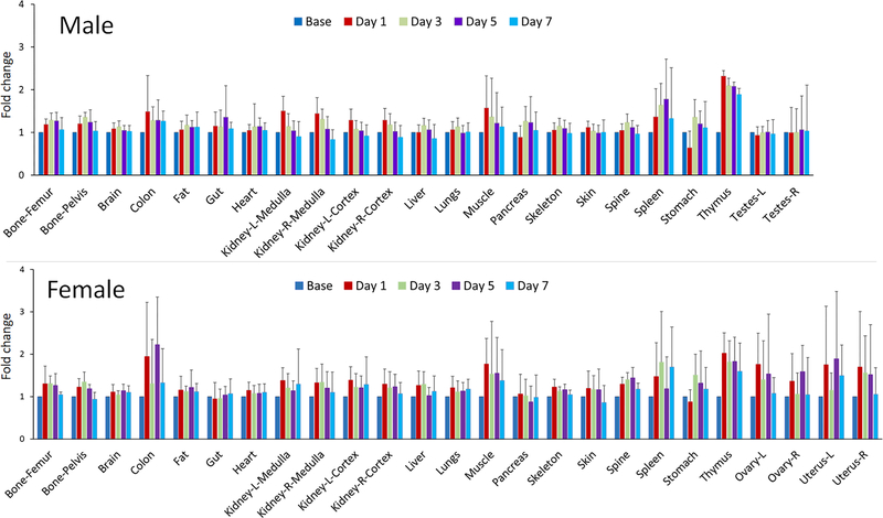
Figure 5.
Relative signal changes in organs/tissues in male and female rats before and at 1, 3, 5 and 7 days after cyclophosphamide treatment (80 mg/kg, single dose). The data are presented as fold change, where the baseline signal intensity in each tissue before cyclophosphamide administration was normalized to 1. Note that the toxicity profiles between the two sexes were visually comparable, where the main susceptible tissues, including the bones, brain, colon, fat, heart, kidneys, liver, stomach and thymus were consistently identified between males and females. Signal changes in the ovaries were also significant as a result of cyclophosphamide treatment.

