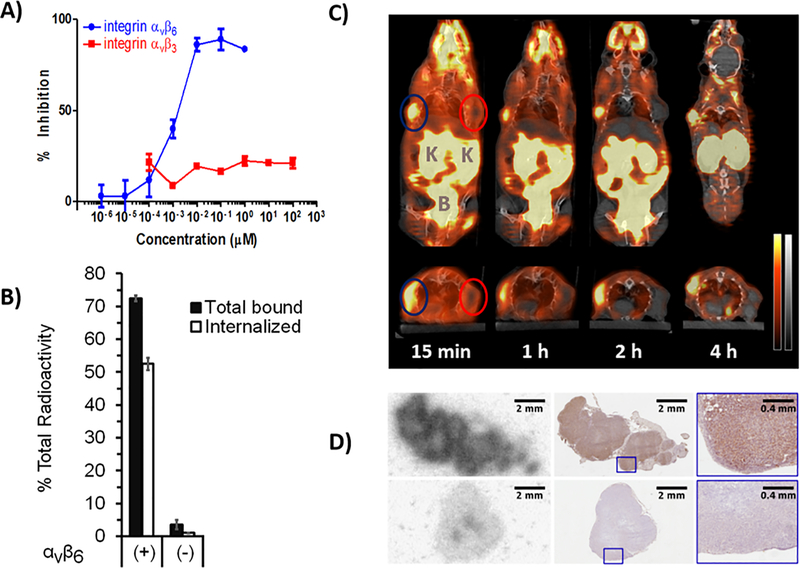
Fig. 1. In vitro affinity and selectivity studies and in vivo mouse studies with [18F]αvβ6-BP. A).
ELISA binding curves of [18F]αvβ6-BP for integrins αvβ6 and αvβ3 against biotinylated fibronectin and vitronectin, respectively (n = 3/integrin/concentration; bars: SD). B) Cell-binding and internalization for [18F]αvβ6-BP using the paired integrin αvβ6-expressing DX3puroβ6 cells(+) and αvβ6-null DX3puro control (−). Filled columns: fraction of total radioactivity (n = 4/cell line/condition; 60 min); bars: SD. C) Representative coronal (upper) and transaxial (lower) cross-sections of PET/CT images obtained after injection of [18F]αvβ6-BP (10 MBq) in mouse bearing paired DX3puroβ6/DX3puro xenografts (112 and 225 mg, blue and red ellipses, respectively). PET: red, CT: gray, K = Kidneys, B = Bladder. D) Autoradiography image of DX3puroβ6 (top) and DX3puro (bottom) tumors harvested 1 h after injection of [18F]αvβ6-BP (37 MBq; left) and matched adjacent immunohistochemistry sections stained for integrin αvβ6 expression (middle, right: magnified sections).

