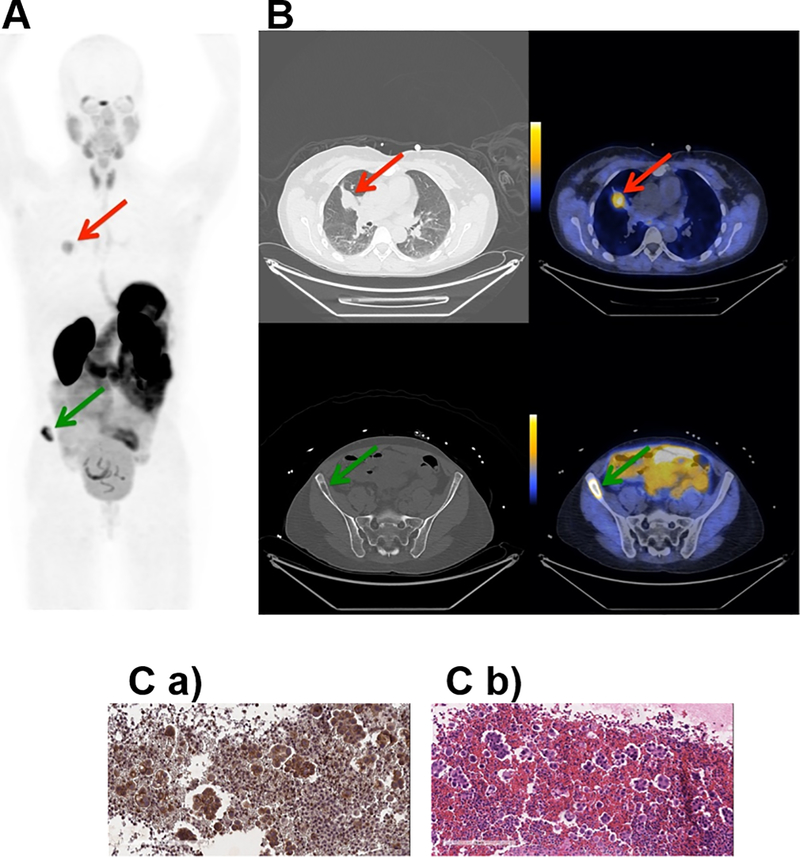
Fig. 2. Representative PET, CT and PET/CT images and IHC staining for subject 1.
Subject 1 was a 53 year old female never-smoker with no significant past medical history diagnosed 20 months prior to study enrollment with stage IV adenocarcinoma of the lung. A. Coronal maximum intensity projection PET image [scaled to SUVmax 15.0] shows distribution of [18F]αvβ6-BP 1 hour after intravenous administration. Red arrow indicates uptake of [18F]αvβ6-BP in primary lung lesion (SUVmax 5.2) and green arrow in the Right iliac wing metastasis (SUVmax 13.5). B. Corresponding axial CT (left) and PET/CT (right) images [scaled to SUVmax 7.0] show distribution of [18F]αvβ6-BP in lung mass (top) and right Iliac bone metastasis (bottom). C. a) Immunohistochemistry section of sample obtained from pleural fluid (no tissue available for the primary tumor or the right iliac wing metastasis) stained for integrin αvβ6-expression and b) corresponding H&E staining.

