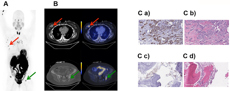
Fig. 3. Representative PET and PET/CT images, and IHC staining for subject 2.
Subject 2 was a 59 year old female diagnosed with Stage IV invasive mammary carcinoma. A. Coronal maximum intensity projection PET image [scaled to SUVmax 15] shows distribution of [18F]αvβ6-BP 1 hour after intravenous administration. Red arrow indicates uptake of [18F]αvβ6-BP in primary breast lesion and green arrow in the left iliac metastasis. B. Corresponding axial CT (left) and PET/CT (right) images [scaled to SUVmax 7] show distribution of [18F]αvβ6-BP in breast mass (SUVmax 3.9) and left Iliac bone metastasis (SUVmax 13.1). C. Immunohistochemistry section stained for integrin αvβ6-expression and corresponding H&E staining of primary breast tumor (a and b) respectively and left Iliac metastasis (c and d) respectively.

