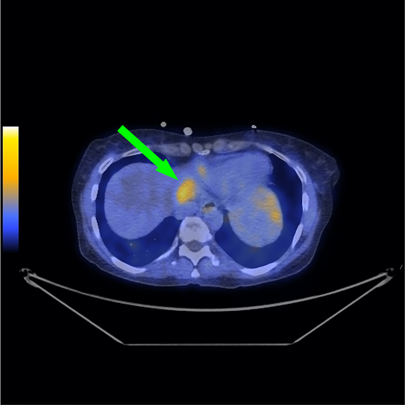
Fig. 5. Representative fused PET/CT for subject 4.
Subject 4 was a 51 year old female diagnosed with initial stage IV adenocarcinoma of the colon with metastases to liver, lungs and abdominal lymph nodes at time of diagnosis. [18F]αvβ6-BP PET/CT images of the upper liver demonstrate elevated activity in the upper left hepatic lobe [scaled to SUVmax 3.0] (green arrow).

