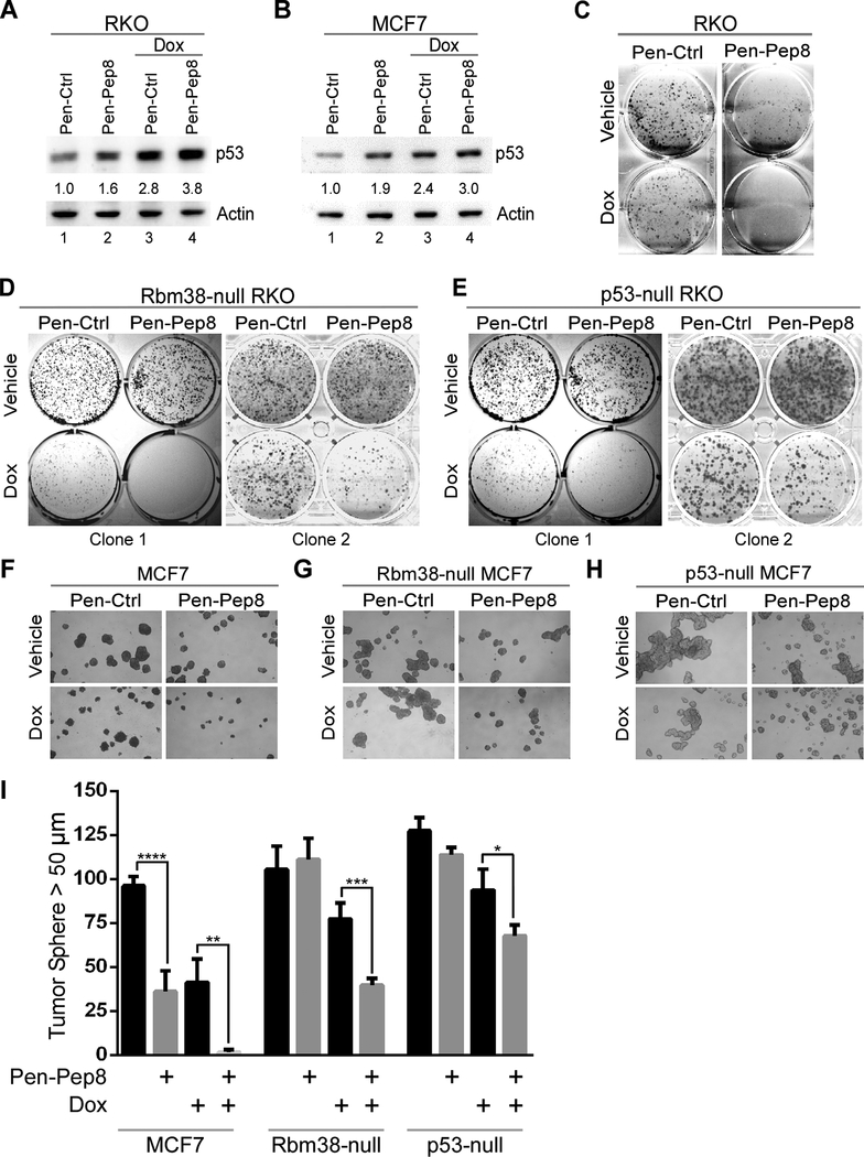
Figure 4.
Pep8 sensitizes tumor cells to doxorubicin. A Immunoblot for RKO cells treated with peptide alone (Pen-Ctrl or Pen-Pep8, 2.5 μM), or in combination with 6.25 ng/mL doxorubicin for 18 h. Standard deviation of relative p53 band intensities was 0.05 (Pep8), 0.7 (Ctrl + Dox) and 0.8 (Pep8 + Dox). B The experiment with MCF7 cells was performed as in A. Standard deviation of relative p53 band intensities was 0.09 (Pep8), 0.2 (Ctrl + Dox) and 0.5 (Pep8 + Dox). C Colony formation assay for RKO cells treated with 5 μM Pen-Ctrl or Pen-Pep8 with and without 3.125 ng/mL doxorubicin. D-E Colony formation assay for Rbm38- and p53-null RKO cells treated with Pen-Ctrl or Pen-Pep8 with and without 6.25 ng/mL doxorubicin. F-H Tumor sphere assays for wild-type, Rbm38- and p53-null MCF7 cells treated with Pen-Ctrl or Pen-Pep8 (5 μM) with and without 6.25 ng/mL doxorubicin. Tumor spheres (> 50 μm) counted after 7 days of treatment with peptides alone or in combination with doxorubicin. I Quantification of tumor spheres from F-H. Values represent the mean ± SEM of three independent experiments (* p=0.028, ** p=0.0068, *** p=0.0028, **** p=0.0013). All western blot figures are representative data of at least 2 independent replicates.

